Copyright © 2024 The President and Fellows of Harvard College | Accessibility | Digital Accessibility | Report Copyright Infringement
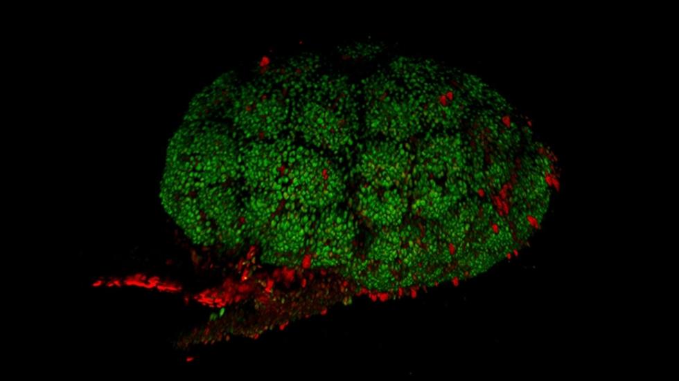
murine embryonic kidney, Leif Oxburgh
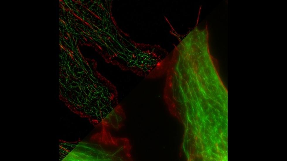
bovine endothelial cells, Doug Richardson/HCBI
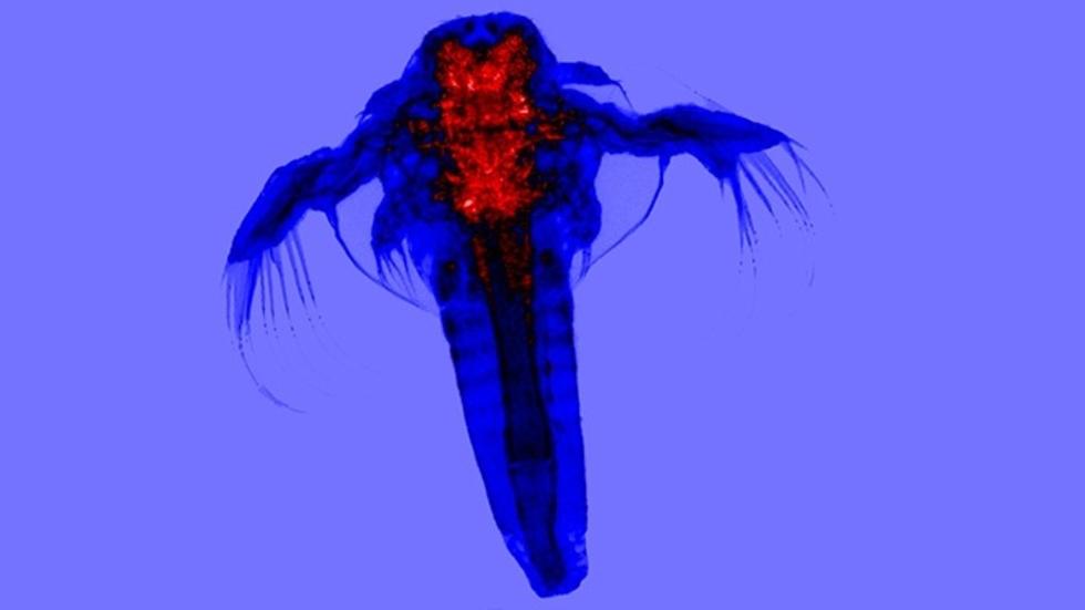
brine shrimp, Casey Kraft/HCBI
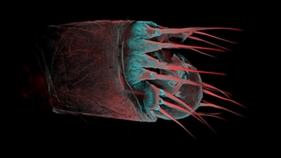
spermatopositor, Taras Dreszer/Giribet lab
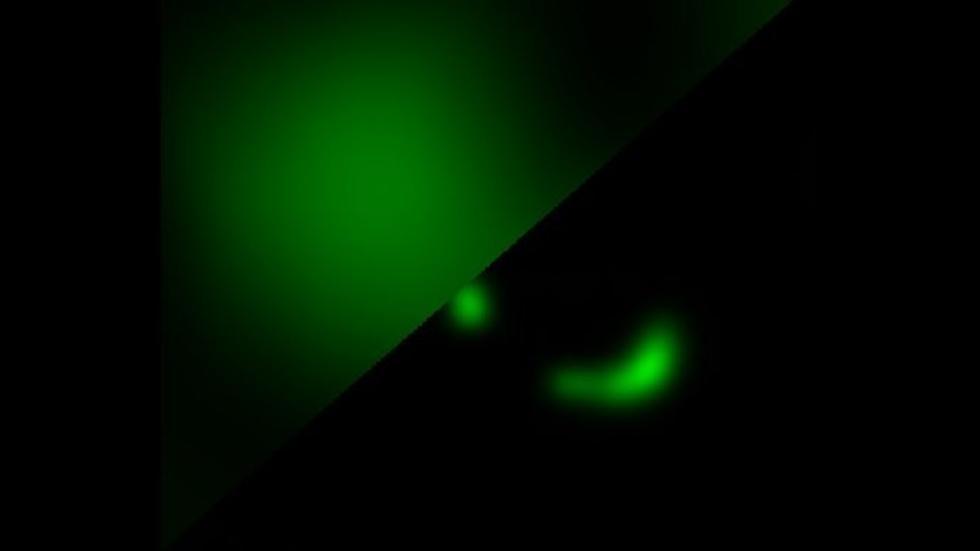
HeLa cell focal adhesion, Doug Richardson/HCBI
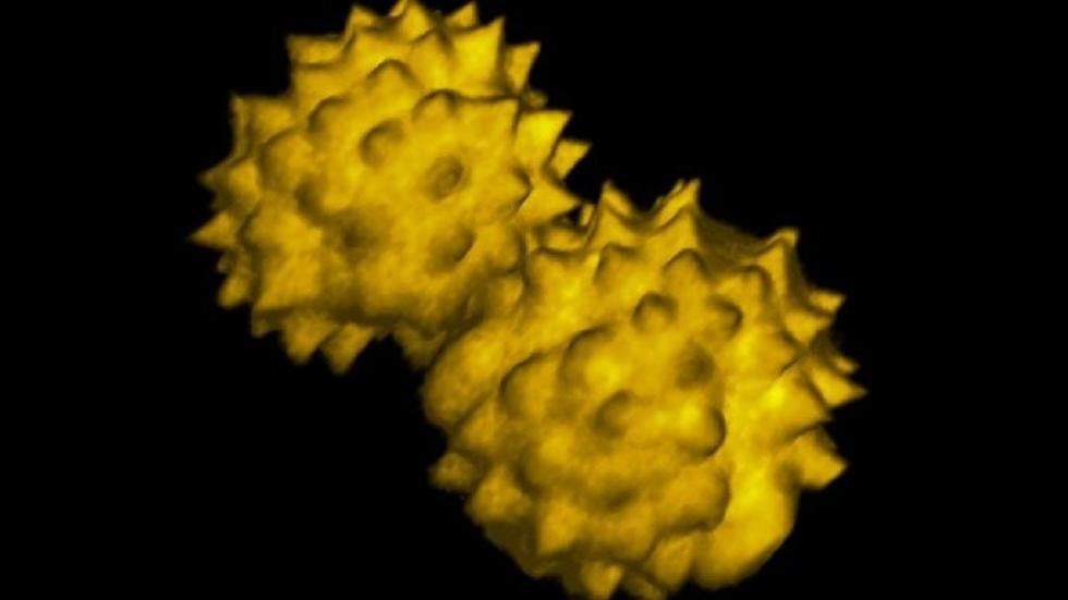
pollen grains, Doug Richardson/HCBI
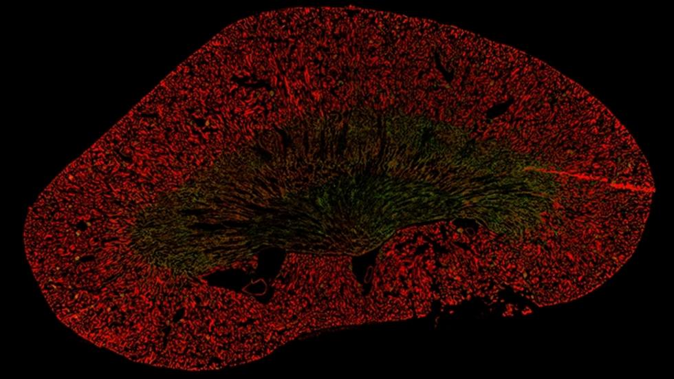
200+ tiled murine kidney, Doug Richardson/HCBI
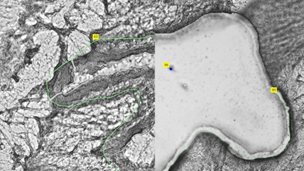
human lung tissue section, Amy Colby/Kobzik lab
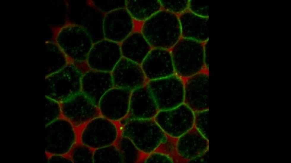
zebrafish embryo, Andi Pauli/Schier lab
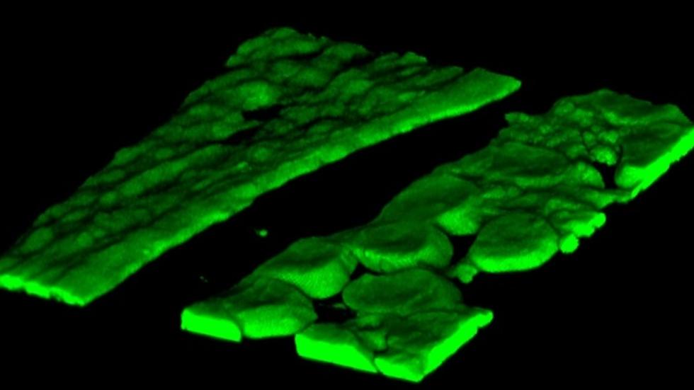
murine kidney section, Doug Richardson/HCBI
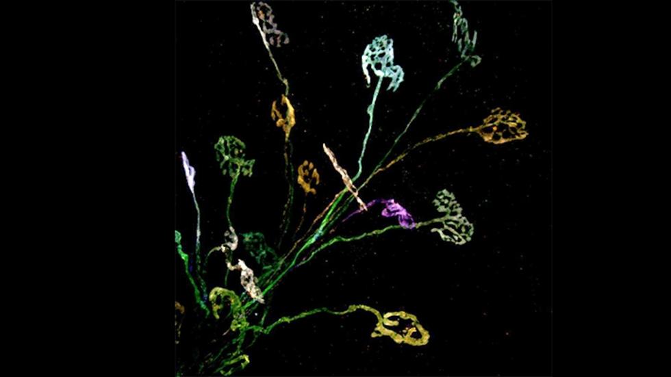
murine motor neurons, Ian Boothby/Lichtman lab, Doug Richardson/HCBI

2014 Recipient
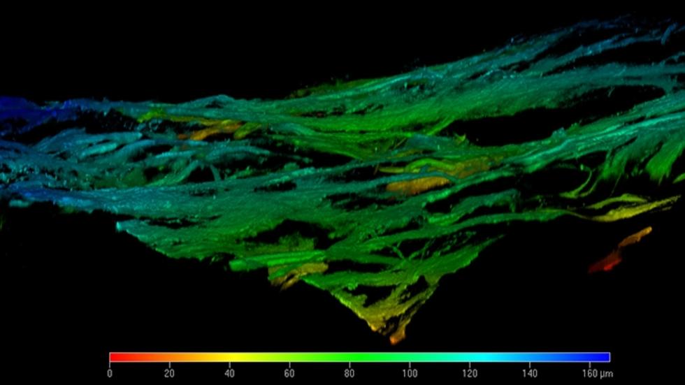
3D collagen matrix, second harmonic generation, Doug Richardson/HCBI
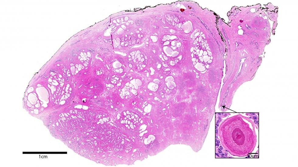
1200+ tiled H&E stained tissue section, Doug Richardson/HCBI
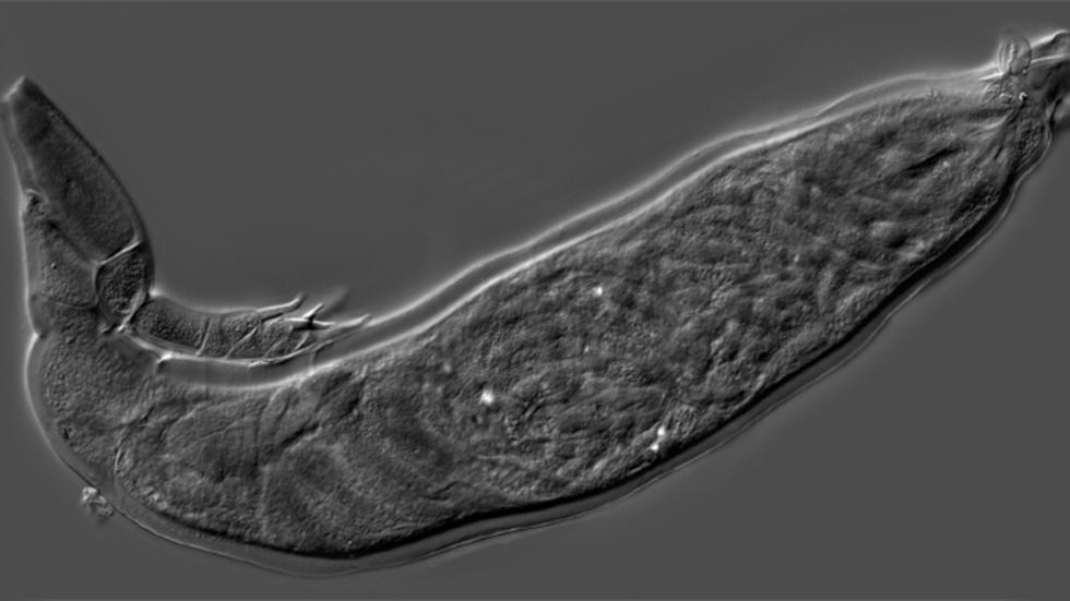
DIC image of H. virescens, Danny Haelewaters/Pfister lab
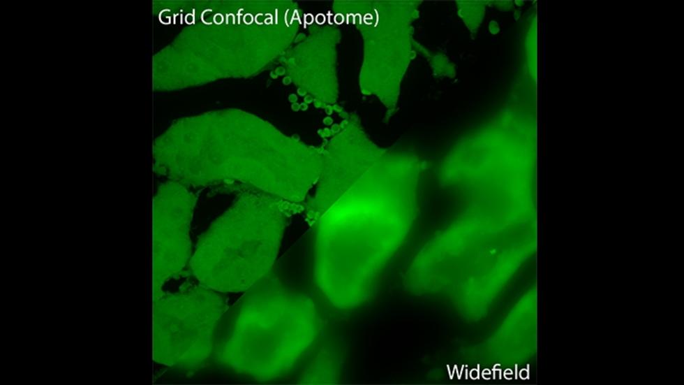
16um mouse kidney, Doug Richardson/HCBI
Room 2052 Biological Labs
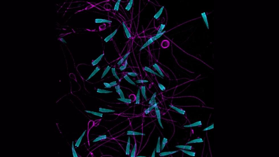
sea urchin sperm, Rick Ng, Megan Parsons, Ellery Jones/MCB 68
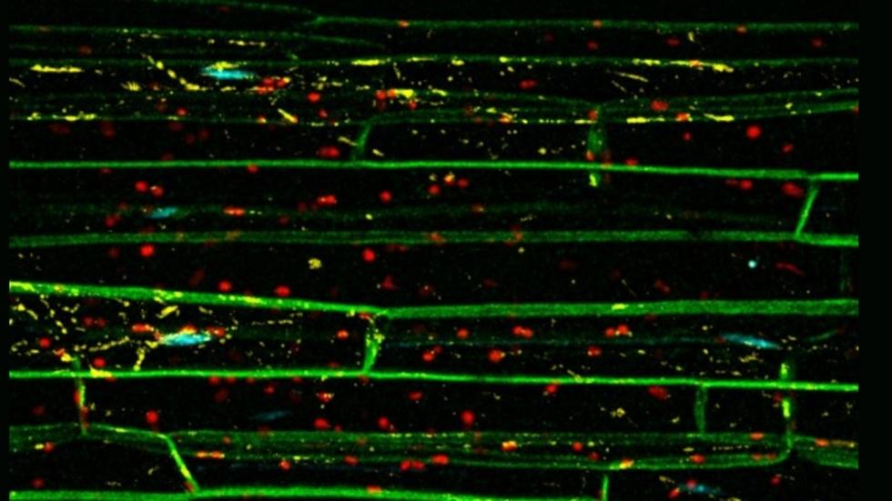
kaleidocell, Karagen Hudson Audrey Chen/MCB 68
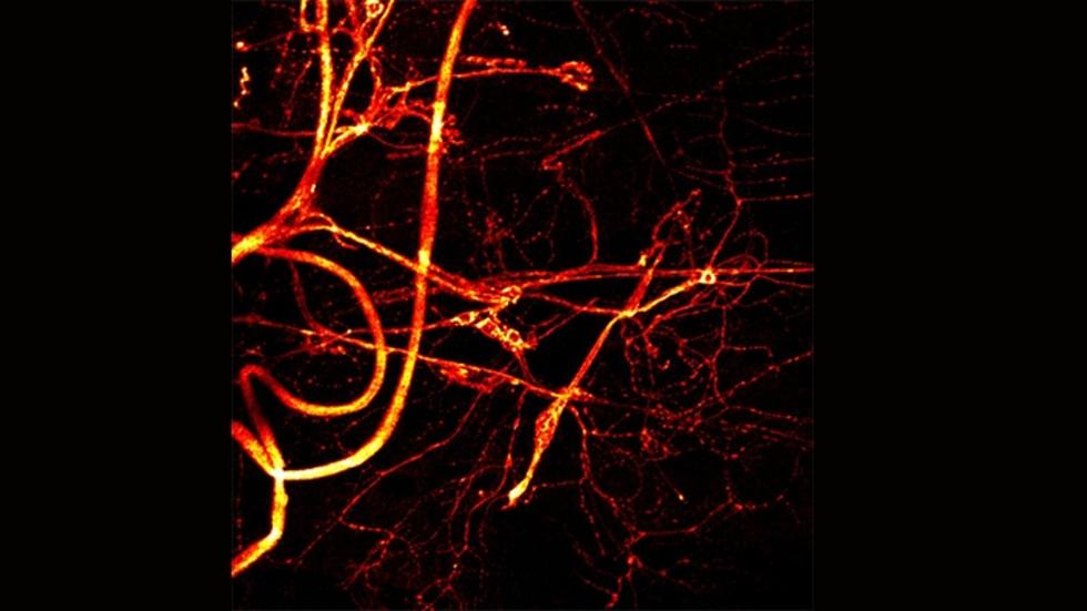
drosophila neuro-muscular junction, Chloe Warinner Jumai Yusuf/MCB 68
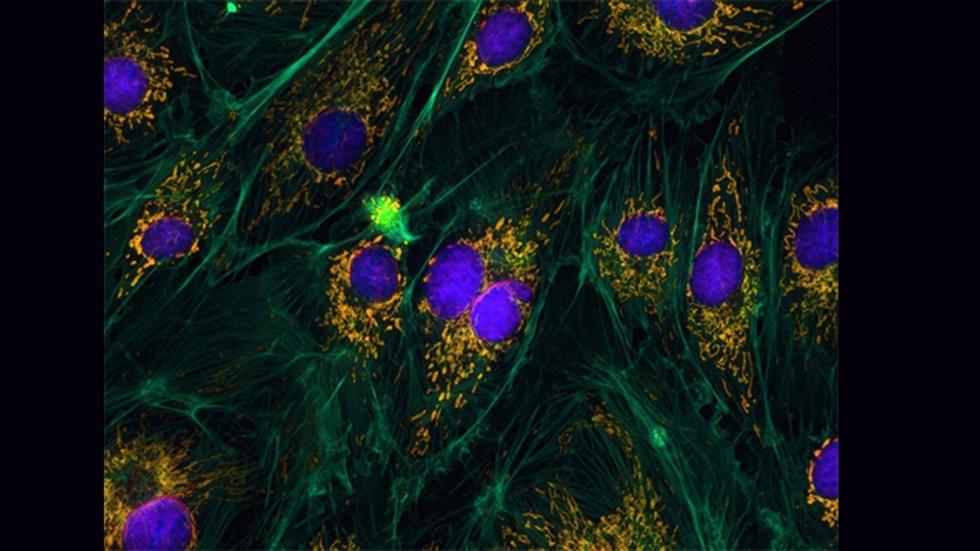
bovine endothelial cells, Bianca Mulaney/MCB 68
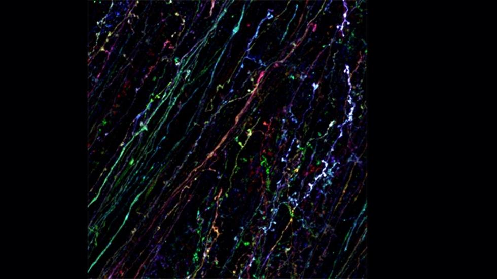
murine brain tissue, Dabin Hwang/MCB 68
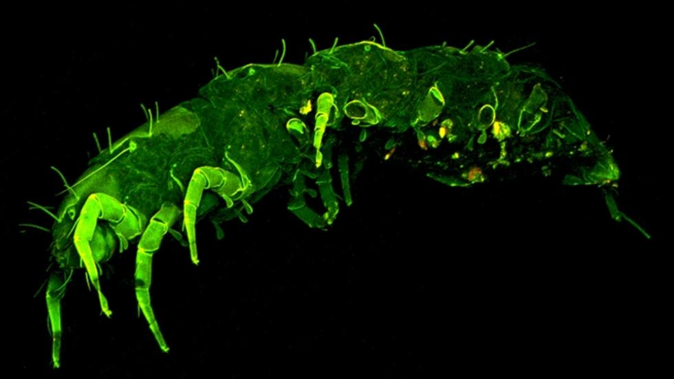
Pauropoda, Georgia Stirtz, Matt Miller, Manny Alvarez Romero, Erik Owen/MCB68
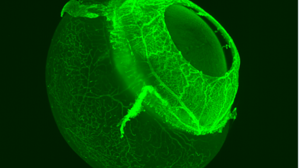
vasculature of the eye, Claudia Prahst/Bentley Lab
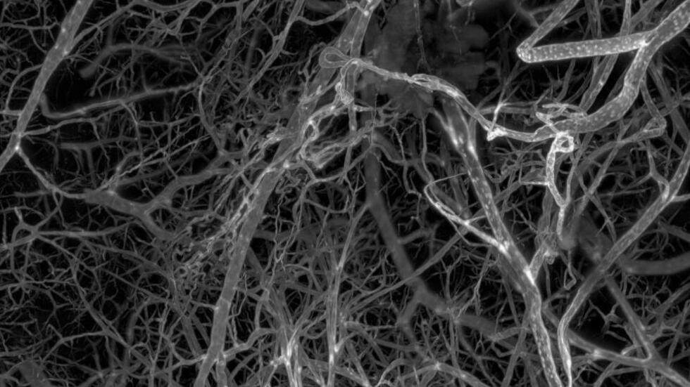
vasculature of the mouse brain, Erin Diel/HCBI
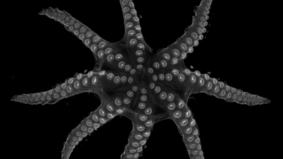
Octopus, Cory Allard, Bellono Lab
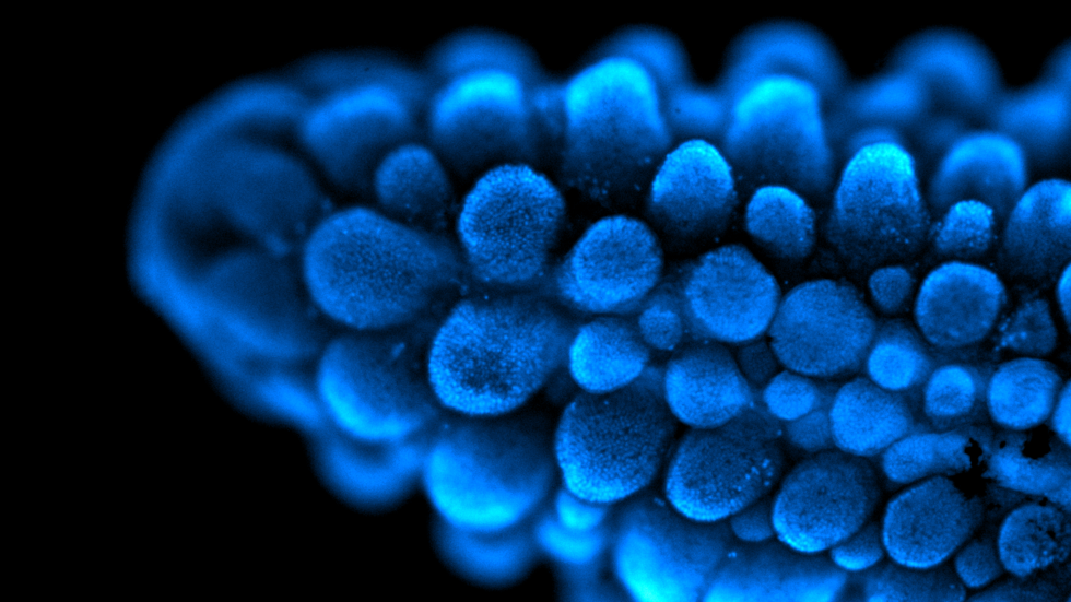
Sea Robin leg, Corry Allard, Bellono Lab
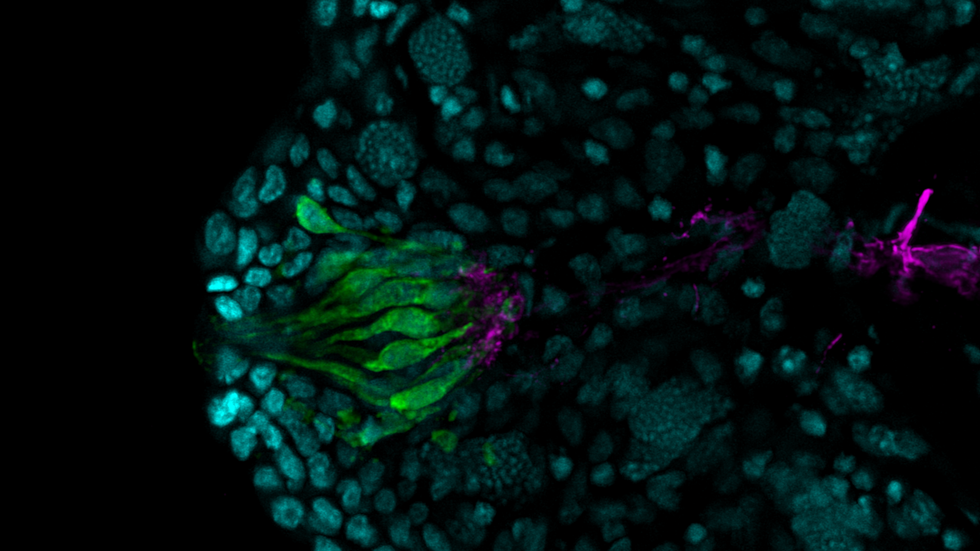
Taste bud, Corry Allard, Bellono Lab
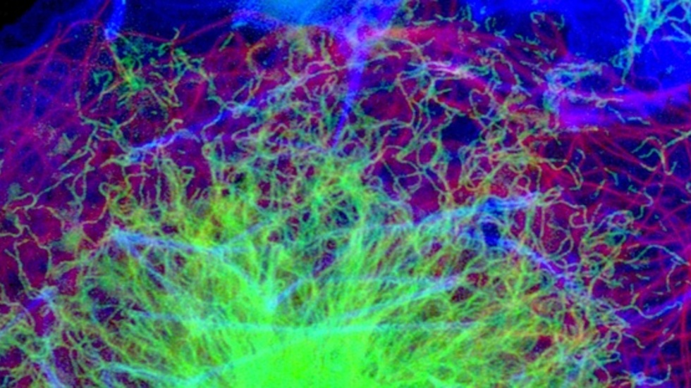
Cytoskeleton of Cos7 Cells, Tommy Krug/ Weitz Lab