Equipment
We have instruments in five locations!
Harvard CBI - Main Lab, 16 Divinity Ave., Cambridge
Live Cell Imaging Core - Satellite Facility, 7 Divinity Ave., Cambridge
Optical Imaging Core - Satellite Facility, 150 Western Ave., Allston
Cryo-EM Center- 11 & 52 Oxford St., Cambridge
Image Data Storage and Analysis - Cloud Infrastructure

Main Facility @ Biological Laboratories, Cambridge

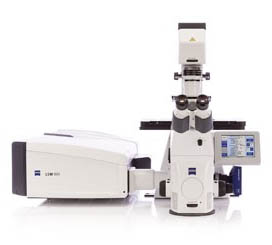
The LSM 980 confocal microscope offers 34-channels of supreme sensitivity, owing to its GaAsP and Airyscan2 detectors. This extraordinary point scanner can simultaneously image and spectrally separate up to 10 different dyes and create high-resolution 3D images over large tiled areas. The unparalleled sensitivity of this system also makes it the ideal live-cell imaging point scanner, allowing for high-speed confocal acquisition with brilliant signal-to-noise ratios. Along with deconvolution, Airyscan2 provides a 1.7x improvement over the theoretical diffraction limit. This system also utilizes the Airyscan2 detector for Zeiss Dynamic Profiler to measure molecular concentration, asymmetric diffusion, and flow dynamics of fluorescent proteins in your living samples.
Laser lines: 405, 445, 488, 514, 561, 594, 640, 761
Commonly imaged dyes: all visible spectrum and near IR dyes (405-750nm)

Multi-photon illumination has always appealed to biologists for imaging deep into living tissue. But did you ever imagine probing more than 5mm into your sample? Using the 980 NLO with our specialized Clearing objective, you can visualize deep into clarified tissue with incredible resolution. Even without clarifying your samples, this microscope excels at penetrating deep into live or fixed specimens, and creating beautiful 3D datasets thanks to a tunable infra-red laser that can be used for multi-photon (2P, 3P) and second harmonic generation (SHG) imaging.
Laser lines (confocal): 405, 440, 458, 488, 514, 561, 633
Laser lines (2-photon): Spectra-Physics Insight DS+ tunable from 680-1300nm
Commonly imaged dyes: all visible spectrum and near IR dyes (405-700nm)
Confocal detection: 6 channel spectral detector
2-photon detection: 6 channel spectral detector or 4 channel (Blue, Green, Red, Far-Red) non-descanned detection
Specification of the LSM 980 NLO
The Nikon Super Resolution Spinning Disk Confocal (SoRa) is a fully-loaded live cell imaging platform with dual disks (SoRa 50 μm) for super resolution and standard confocal imaging, Hamamatsu Quest 2 qCMOS & Andor iXon 888 EMCCD and, full incubation. The system is capable of up to 120nm resolution with the SoRa disk and 10s of nm with SMLM. DNA-paint and some dyes are possible for SMLM, but please speak to the imaging team prior to your experiment.
Laser Lines: 405,488,561,640
Objective magnifications 4x/0.2, 10x/0.4,, 25x/1.05, 40x/1.15, 63x/1.42, 63x/1.49 TIRF
The LSM 910 is a high-performance laser scanning confocal microscope designed for flexible and sensitive imaging of fixed and live samples. This system is well-suited for users requiring high-quality imaging of cellular structures, tissue sections, and complex biological samples. With Lightfield 4D, you no longer have to compromise, as you can capture 80 volumes per second without time delay in 3D. This makes it possible to follow neuronal activity in zebrafish brains, track tissue movement in developing Drosophila embryos, and keep track of moving structures in crawling C. elegans larvae. The unique one-snap-one-volume imaging ensures that crucial events are not missed or distorted.
Laser lines: 405, 488, 561, 640
Commonly imaged dyes: DAPI, Alexa 488/GFP, Alexa 555/568/tdTomato, Alexa 647
LSM 910 Lightfield 4D Specifications
The LSM 900 laser scanning confocal microscope is optimal for confocal imaging with the four commonly used dyes. The upright configuration is perfect for imaging organisms such as zebrafish. The entire system is fully motorized for autofocusing over large fields of view. But did you ever imagine probing more than 5mm into your sample? Using the LSM 900 with our specialized Clearing objective, you can visualize deep into clarified tissue with incredible resolution. The system has 2 PMTS, a GaSaP detector, and a T-PMT for transmitted light.
Laser lines: 405, 488, 561, 640
Commonly imaged dyes: DAPI, Alexa 488/GFP, Alexa 555/568/tdTomato, Alexa 647
A top of the line fully spectral point scanning workhorse, the LSM 880 can handle all of your fixed-sample imaging demands. With a 440nm laser line, this confocal is particularly suited to imaging "cyan" dyes and fluorescent proteins. It is also equipped with pulsed 405 and 440 lasers and a 2-channel Big.2 detector for performing Fluorescence Lifetime Imaging (FLIM) measurements. The system is also equipped for imaging single molecules through fluorescence correlation techniques.
Laser lines: 405, 440, 458, 488, 514, 561, 594, 633
Commonly imaged dyes: all visible spectrum and near IR dyes (405-700nm)

So you have a four-color sample that requires 3D imaging over large tiled areas? Then the LSM 700 is the microscope for you. This incredibly user-friendly point scanner can image up to two channels simultaneously, due to its unique Variable Secondary Dichroic, all the while acquiring 3D images over multiple fields of view. In addition, the LSM 700 boasts a back-thinned Hamatsu EMCCD camera for performing high-sensitivity wide-filed imaging.
Laser lines: 405, 488, 555, 638
Commonly imaged dyes: DAPI/BFP, GFP/488, 555/568/tdTomato, 647

Long-term,high-speed 3D imaging with virtually no phototoxicity or bleaching may sound too good to be true, but the Lightsheet.7 delivers. Capable of imaging both aqueous (RI=1.33) and cleared tissue (RI=1.45) samples, this revolutionary microscope acquires 3D images that previously took hours on a point scanner in mere minutes! The ability to freely rotate your sample and image from multiple angles (multi-view), improves resolution and imaging depth.
Laser lines: 405, 488, 561, and 638
Commonly imaged dyes: DAPI. BFP, GFP, 488, tdTomato, dsRed, 555, 568, 647
RI=1.33 optics: 5x/0.16, 20x/1.0, 40x/1.0
RI=1.45 optics: 5x/0.16, 20x/1.0 (5.5mm working distance)

Have you ever looked at a giant pile of slides, and wondered how you'll find the time to image them all? The Axio Scan.Z1 is your answer! This high speed slide scanner has an astonishing 100-slide capacity, and can image samples stained with both colormetric and fluorescent dyes. This particular system is configured for maximum color seperation, an additional infra-red imaging channel and the ability to perform polarized light imaging as well.
Cameras: AxioCam 705 Color & Hamamatsu Orca Flash 4.0
Transmitted Light: Brightfield, TIE contrast, Circular-Polarization
Filter sets: quadruple bandpass (Blue, Green, Red, Far-Red), DAPI/BFP, 488/GFP, 555/568/tdTomato, 594/mCherry, 647/Cy5, 750/Cy7
Best dye combinations: DAPI/488/568/647/750

The MERSCOPE combines MERFISH technology with high resolution imaging, fluidics, image processing, and automation to deliver a complete end-to-end spatial genomics solution. The MERSCOPE Platform comes with everything you need to easily acquire and analyze high-quality MERFISH measurements.

Widefield, laser widefield, TIRF, and super-resolution? The ELYRA can do it all! Using lattice structured illumination, this system can improve resolution in X, Y, and Z two-fold – with no special sample prep needed. The lattice illumination pattern is especially gentle for live-cell imaging. If that isn’t sufficient for your experiment, single molecule localization (SML) super-resolution techniques like PALM or dSTORM can improve your resolution by as much as an order of magnitude.
Laser lines SIM/PALM/STORM: 405, 488, 561, and 638
Commonly imaged dyes SIM/PALM/STORM: DAPI. BFP, GFP, 488, tdTomato, dsRed, 555, 568, 647

Through the combination of proprietary Yokogawa high-speed Confocal Scanner Technology, water immersion lenses, up to four high field-of-vision cameras, a microscopic stage with cell cultivation environment, and an integrated robotic pipetter, the unit provides high throughput, high-resolution imaging, and phenotypic screening, making it the ideal choice for a variety of applications.
Laser Lines: 405, 445, 488, 561, 640
Commonly Imaged Dyes: DAPI, Alexa 488/GFP, YFP, Alexa 555/568/tdTomato, Alexa 647
The CellVoyager High-Content Analysis System CQ3000 is a high-content analysis platform that rapidly acquires high-resolution 3D images. It enables advanced assays in environments closer to the human body, such as organoids—which replicate certain structures and functions of human organs— or organ-on-a-chip models that mimic physiological functions of organs on microfluidic chips. In addition, multicolor imaging with multiple fluorescent dyes enables complex assays, such as morphological profiling of cellular components and organelles, using techniques such as Cell Painting.
Laser Lines: 405, 488, 561, 640
Commonly Imaged Dyes: DAPI, Alexa 488/GFP, Alexa 555/568/tdTomato, Alexa 647

The STEDYCON is a confocal microscope and STED nanoscope with a resolution down to 30 nm. With its super-intuitive user interface, the STEDYCON provides an intelligent microscope platform that enables everyone to acquire superb superresolution images. Define your experiment and enjoy fine imaging at the push of a button!
Excitation Laser Lines: 405, 488, 561, 640
Depletion Laser Line: 775
Objectives: 10x/0.3, 20x/0.5, 100x/1.46
Common Confocal Dyes: DAPI/488/568/647
Common STED Dyes: STAR RED, ATTO647N, JF646, STAR ORANGE, AF594, ATTO590

Combining a 16x zoom with high numerical aperture objectives, the AxioZoom.V16 offers superior brightness in large object fields. This highly versatile microscope can image samples illuminated by transmitted, reflected, polarized, and fluorescent light sources. It is equipped with a high resolution color camera and a photometrics Prime 95B back-illuminated sCMOS camera for fast, highly sensitive imaging.
Cameras: Prime 95B BI, Axiocam 712 color
Filter sets: DAPI, CFP, GFP, YFP, RFP, mCherry, Far Red
Best dye combinations: DAPI/BFP, GFP/488, tdTomato/dsRed/mCherry/555/568/594, 647
or CFP, YFP, tdTomato/dsRed/mCherry/555/568/594/647

Even the world’s greatest surgeon might struggle with dissecting single cells out of a slice of tissue. With the PALM system, laser microdissection, catapult and capture are as easy as drawing a region to dissect on the screen, and pressing start. Remove individual cells from your sample slices, or excise entire subregions for RT-PCR, DNA, or even mass spec analysis.
Laser lines: 350
Filtersets: DAPI, GFP, RFP

Are you tired of waiting for your tissues to passively clear? Speed up the Process with the X-CLARITY Tissue Clearing System. Perform the lipid removal step of the CLARITY protocol in as little as 3 hours (2mm slice) or 24 hours (Whole brain). ETC buffer is included with your reservation!
HCBI CLARITY protocol
X-CLARITY User Guide
Live Cell Imaging Core @ Sherman Fairchild, Cambridge

The Bruker TruLive 3D system is designed for long-term,high-speed 3D imaging of live or fixed tissue. This system only allows for imaging of samples in aqueous based media so it is unable to accomodate any cleared tissue sampels. The system utilizes a unique trough samples holder design, allowing or visualization of samples without the need to embed them in agar or other restraining media.
Laser Lines: 405,488,561,642
Objective magnifications 12.5x, 25x, 37.5x, 50x

The Nikon Spinning Disk Confocal (CSU-W1) is a fully-loaded live cell imaging platform with dual disks (25 and 50 um) for low and high magnification imaging, dual Photometrics BSI cameras, full incubation, and a programable 405 nm photoconversion/optogenetics laser. This system has full incubation and is ready to image your live samples at top speed.
Laser Lines: 405,488,561,594,640
Photomanipulation: 405, 473
Objective magnifications 4x/0.2, 10x/0.4, 20x/0.8, 25x/1.05, 63x/1.42
Optical Imaging Core @ Science and Engineering Complex, Allston
A member of the HCBI training team is on site Monday & Wednesday 9am-5pm

This LSM 900 comes fully loaded for all of your bioengineering needs. A Hamamatsu Flash 4.0 camera allows for fast, sensitive, widefield fluorescence imaging. The confocal scan head can perform standard multicolor confocal imaging and spectral imaging and create patterned excitation for FRAP/optogenetics experiments. Additionally, an Airyscan 2.0 detector is available to improve resolution beyond the diffraction barrier and increase confocal sensitivity. The entire system is fully motorized, allowing autofocus over large fields of view. Incubation and environmental control will keep your samples happy for the duration of the experiment.
Objectives: 2.5x/0.085, 10x/0.45 air, 20x/0.8 air, 40x/1.2 water, 40x/1.3 oil
Laser lines: 405, 488, 561, 640
Commonly imaged dyes: DAPI, Alexa 488/GFP, Alexa 555/568/tdTomato, Alexa 647
This LSM 900 confocal is configured to be a materials workhorse. It comes equipped with black and white and color cameras that can image a wide range of widefield contrasts: DIC, phase contrast, polarization, reflected light, and fluorescence. The confocal scan head can image in fluorescence or reflected light mode.
Objectives: 2.5x/0.085 air, 5x/0.3 air, 10x/0.25 air, 20x/0.5 water dipping ,50x/0.55 air,
Laser lines: 405, 488, 561, 640
Commonly imaged dyes: DAPI, Alexa 488/GFP, Alexa 555/568/tdTomato, Alexa 647
Have you ever looked at a giant pile of slides, and wondered how you'll find the time to image them all? The Axio Scan.Z1 is your answer! This high speed slide scanner has an astonishing 100-slide capacity, and can image samples stained with both colormetric and fluorescent dyes. This particular system is configured with the ability to perform polarized light imaging as well.
LED excitation: 385, 430, 475, 511, 555, 590, 630 nm
Objectives: 5x/0.16 air, 10x/0.45 air, 20x/0.8 air
Filter sets: quadruple bandpass (Blue, Green, Red, Far-Red), DAPI/BFP, 488/GFP, 555/568/tdTomato, 594/mCherry, 647/Cy5
Best dye combinations: DAPI/488/568/647
Cryo-EM Center @ LISE & Northwest Building, Cambridge
The facility is open 24/7 with card access.
A member of the HCBI training team can be reserved for assistance Monday-Friday 8am-5pm

The Glacios 2 Cryo-Transmission Electron Microscope (cryo-TEM) is a 200 kV microscope that allows you to easily collect near atomic data from a broad range of biological targets. It features an integrated Falcon 4i Direct Electron Detector, X-FEG source, fringe-free imaging, and Selectris energy filter which combine to enhance image quality, automate data acquisition, and simplify your work. The Glacios 2 Cryo-TEM is ideally suited for single-particle analysis, cryo-electron tomography (cryo-ET), and micro-electron diffraction (MicroED) applications.
Location: LISE building, 11 Oxford Street, Cambridge

The Vitrobot Mark IV System offers semi-automated vitrification to provide fast, easy, and reproducible sample preparation for cryo-EM. It performs the cryo-fixation process at constant physical and mechanical conditions like temperature, relative humidity, blotting conditions, and freezing velocity. This ensures high-quality cryo-fixation results and a high sample preparation throughput prior to cryo-TEM observation.
Location: Northwest Building, 52 Oxford Street, Cambridge (Installation in 2026)

This newest version of the Wohlwend Compact has a patented valve system that ensures complete and even freezing of samples up to 500 µm in thickness, more than double other systems on the market. In addition, it is compatible with a wide variety of sample carriers including: 3 mm, 6 mm, copper tubes, omega style carriers, cellulose capillary tubes and sapphire disks.
Location: Northwest Building, 52 Oxford Street, Cambridge

The HCBI offers access to two EM Automated Freeze Substitution (AFS2) systems from Leica (Room B412.60, Northwest Building). The systems allow for manual or automated freeze substitution, progressive lowering of temperature (PLT), low temperature embedding and UV polymerization of resins. Both AFS systems are equipped with a freeze substitution processor (FSP) unit that automatically dispenses reagents for both freeze substitution and PLT applications. The LED illumination from within the chamber and the attached stereomicroscope allow for viewing and positioning of samples to ensure ease of use.
Location: Northwest Building, 52 Oxford Street, Cambridge
Data Storage and Analysis
The HCBI offers access to cloud data storage and virtual workstations for all users in the facility.
Data Storage
The HCBI offers 7 shared storage servers that are available to all registered users (hcbishare 1-7) for data storage. The servers have a storage capacity of nearly 3 Petabytes; however, this is not meant as a place to archive your data permanently. When any server approaches 80% full, we will delete files beginning with the oldest first. Only a single copy of your data is stored on these servers, and it is not backed up in multiple locations. As always, we recommend you keep multiple copies of your data.
When accessing the storage servers make sure you log into the correct server to access data from the instrument you used. The Instruments are distributed across the various servers as follows:
hcbishare1: Lightsheet Z7, Axiozoom
hcbishare2: Axioscan #1, 2-photon, MERSCOPE
hcbishare3: Axioscan #2, CD7, LSM 900 Live Cell
hcbishare4: ELYRA, LSM880 FLIM, STEDYCON, CV8000
hcbishare5: LSM980+Airyscan, LSM900
hcbishare6: Allston-Axioscan, Allston-LSM900-BIO, Allston-LSM900-MAT
hcbishare7: Nikon Spinning Disk, Bruker TruLive
Instructions on how to access these storage servers from a non hcbi computer can be found here
There are 10 virtual workstations available for image analysis, hcbiview01-10. These workstations are directly connected to our shared servers, for easy data access.
Calendars for each computer can be found on SPINAL, and it is required that you book time for them here to make sure you have access to the system and the software you may need for your analysis.
Various software is available on these workstations for your image analysis needs, but not every workstation has a copy of every software.
hcbiview01 is reserved for Imaris users only.
hcbiview02-04 are reserved for Arivis users.
hcbiview05-10 are for Zen image analysis and open source programs such as Fiji/ImageJ and Cell Profiler
hcbiview09-10 are for Nikon elements and the Lux bundle for the Bruker Truelive.
Please use the appropriate workstation for the software you wish to use.
Instructions on how to gain access to these remote workstations can be found here.
Workstation specifications:
8 TB SSD storage (connected to a 3 PT storage server with high speed (25Gbit) network cable/cards)
396GB RAM
NVIDIA RTX 8000 GPU
Dual Intel Xeon 20-core processors
Contact Us
Don't know which instrument is right for you? Email us at hcbi_imaging@fas.harvard.edu